3D Imaging Reveals How T Cells Target Cancer — SkimNews
Get the Health newsletter
Daily health & science — research, biotech, public health, the studies worth knowing. Free.
- Cytotoxic T lymphocytes form an immune synapse to deliver toxic molecules precisely to cancerous or infected cells, minimizing damage to surrounding healthy tissue
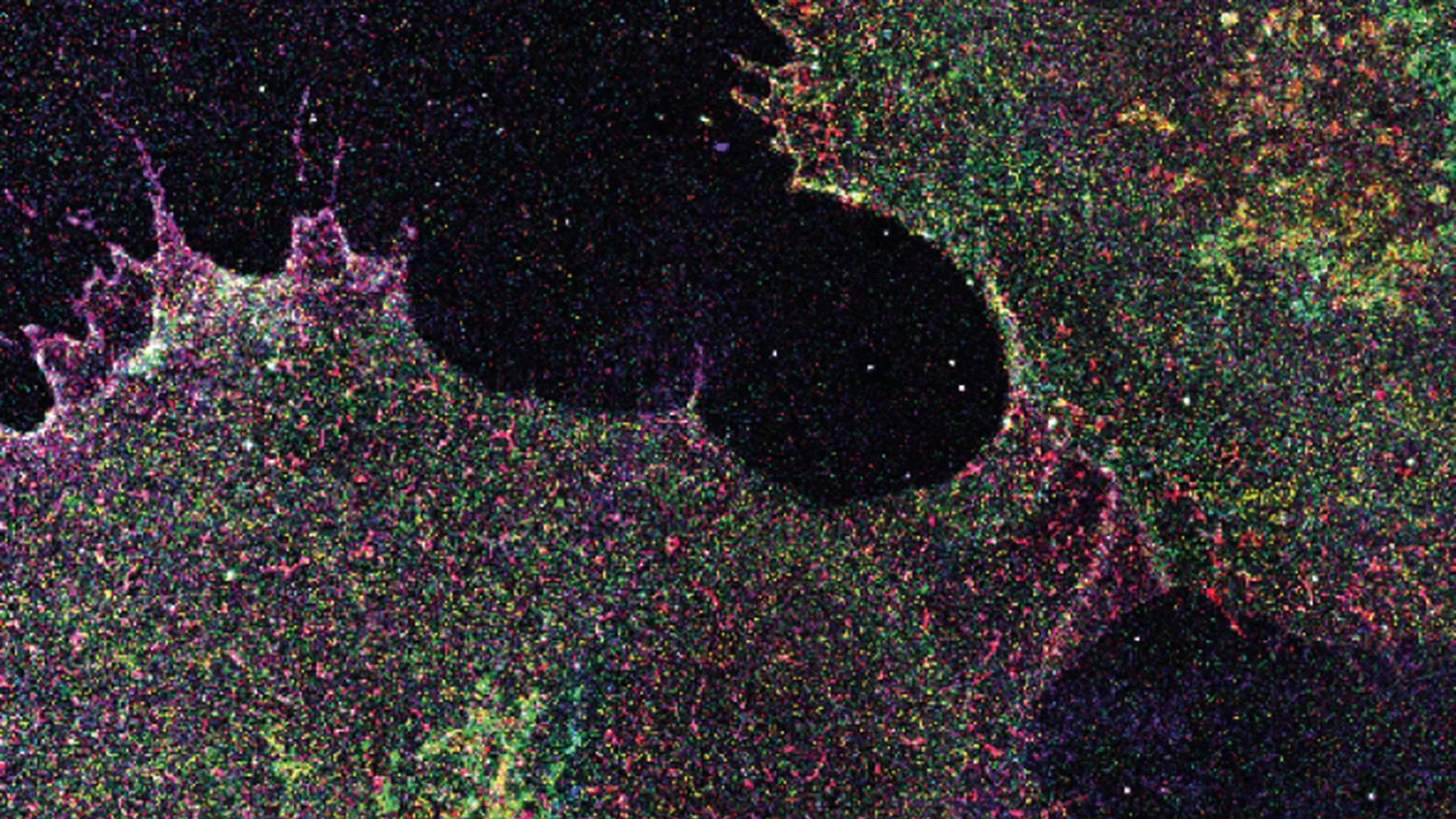
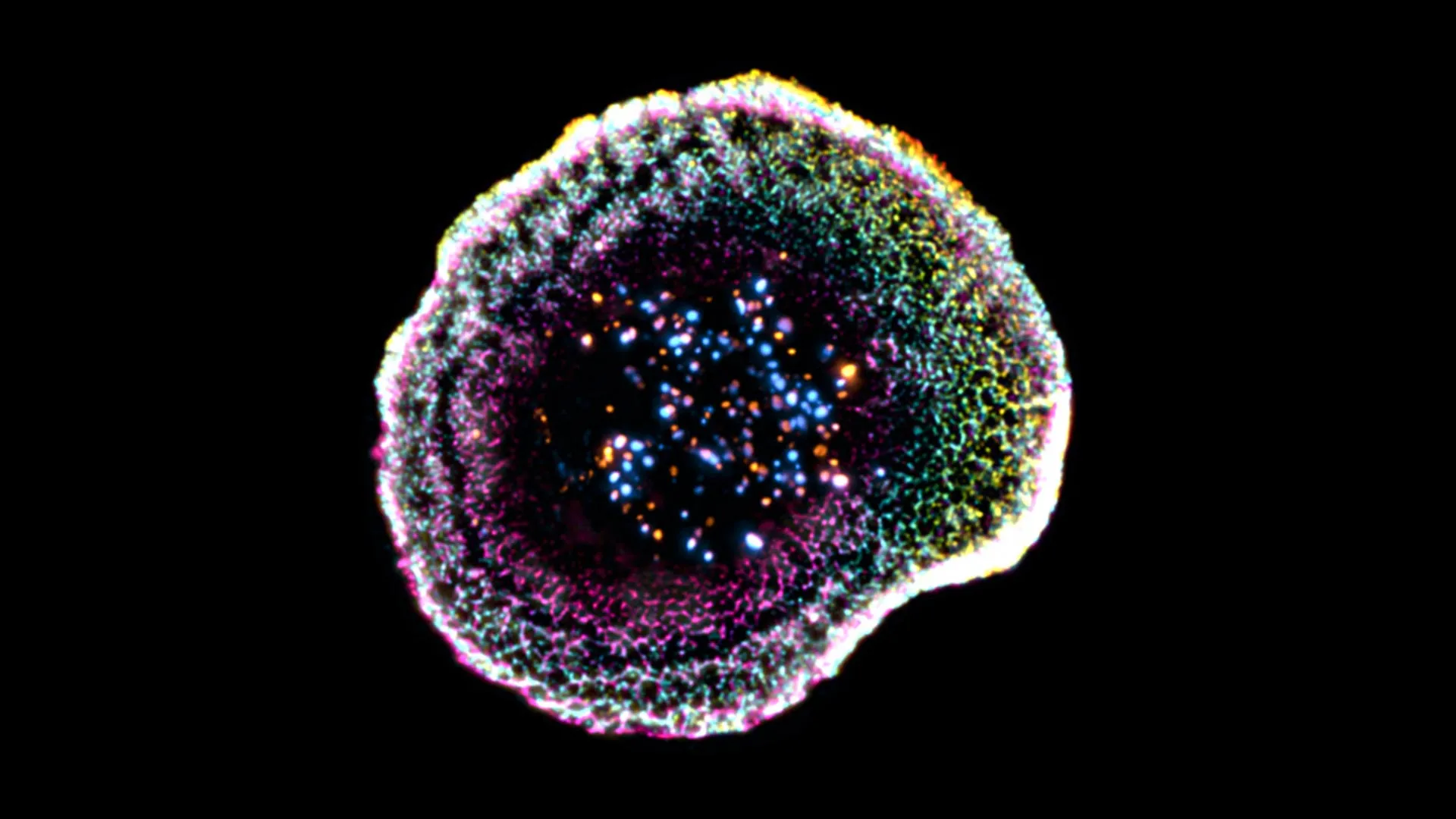
- University of Geneva (UNIGE) and Lausanne University Hospital (CHUV) researchers visualized the internal structure of T cells in 3D under near-native conditions using cryo-expansion microscopy
- Cryo-expansion microscopy (cryo-ExM) preserves cellular architecture by vitrifying cells rapidly and expanding them with a hydrogel, enabling high-resolution, large-scale imaging of intact immune synapses
- Virginie Hamel explains that cryo-ExM overcomes limitations of traditional imaging methods, which often distort delicate biological structures due to sample preparation artifacts
- The study, published in Cell Reports, provides new insights into the nanoscale organization of cytotoxic T cells, advancing understanding of their cancer-killing mechanism
Why it matters: This imaging breakthrough allows researchers to see exactly how T cells target cancer at the molecular level, which could accelerate the development of more effective immunotherapies by improving how we engineer or harness these cells in treatment. The method’s ability to preserve native structure means future studies can rely on more physiologically accurate data.
Ask SkimNews