MIT Maps 3D Structure of Relaxor Ferroelectric — SkimNews
Get the Health newsletter
Daily health & science — research, biotech, public health, the studies worth knowing. Free.
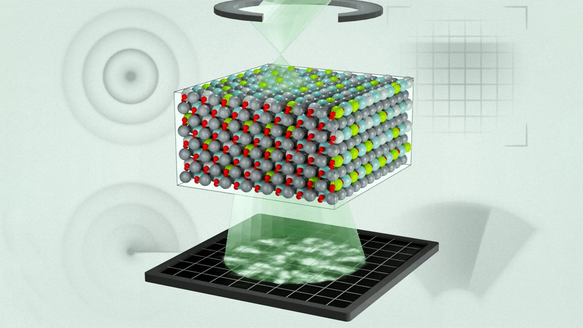
- MIT and collaborators mapped the three‑dimensional atomic structure of a relaxor ferroelectric for the first time using multi‑slice electron ptychography (MEP).
- James LeBeau said the new structural insight will let scientists predict and engineer material properties for computing, energy, and sensor technologies.
- Michael Xu reported that the observed chemical disorder was not fully considered in previous models, prompting refinements.
- Menglin Zhu noted that nanoscale polarization regions were significantly smaller than earlier simulations predicted, leading to updated model predictions.
- U.S. Army Research Laboratory (with the U.S. Office of Naval Research and U.S. Department of War) funded the work, underscoring defense interest in advanced sensor materials.
- Science will publish the study, which shows electron ptychography can directly connect three‑dimensional polar structures with molecular dynamics calculations for the first time.
Why it matters: The atomic‑scale map validates and refines computational models, enabling engineers to design more efficient relaxor ferroelectric sensors, actuators, and energy‑storage devices; defense agencies and commercial manufacturers gain higher‑performance materials while outdated, unvalidated models become obsolete. The breakthrough also showcases multi‑slice electron ptychography as a versatile tool for probing disordered materials, expanding its utility beyond ferroelectrics.
Ask SkimNews


