Cedars‑Sinai Launches 3‑Min MRI to Measure Oxygen — SkimNews

Get the Health newsletter
Daily health & science — research, biotech, public health, the studies worth knowing. Free.
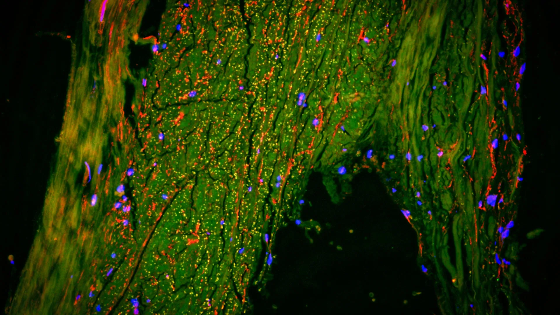
- Cedars‑Sinai Medical Center researchers published a study in Science Translational Medicine showing a three‑minute MRI add‑on can quantify cardiac oxygen consumption without catheters, contrast agents, or breath‑holding.
- The experimental MRI technique was first validated in pigs and then in 22 post‑heart‑attack patients, achieving oxygen‑consumption measurements that matched invasive catheter and PET benchmarks.
- Hsin‑Jung Yang, an associate professor at Cedars‑Sinai, describes the method as a motion‑insensitive, signal‑quantification model that translates blood‑oxygen‑level‑dependent signals into numeric oxygenation values.
- The trial demonstrated that the three‑minute scan can be integrated into routine cardiac MRI protocols, allowing repeated, low‑risk monitoring of heart‑failure patients, especially those with metabolic syndrome.
- GLP‑1 and SGLT2 drugs are now included in clinical guidelines for treating HFpEF, reflecting a shift toward metabolic‑focused therapies for heart‑failure patients.
Why it matters: Patients with heart‑failure, especially those with metabolic syndrome, gain a rapid, low‑risk way to assess cardiac oxygen consumption, while clinicians can replace invasive catheter or PET scans with a three‑minute MRI add‑on, avoiding radiation exposure and contrast agents and enabling more frequent monitoring.
Ask SkimNews