Vitamin A Signals Convert Blue Cones in Fetal Eye — SkimNews
Get the Health newsletter
Daily health & science — research, biotech, public health, the studies worth knowing. Free.
- Johns Hopkins University researchers found that between weeks 10 and 14 of fetal development, blue cone cells in the developing foveola convert into red and green cones, driven by retinoic acid (a vitamin A-derived molecule) and thyroid hormones.
- The finding overturns a ~30-year-old model in which blue cones simply migrated outward from the retina's center; instead, the cells stay in place and change identity, with retinoic acid cutting off new blue cone formation first, then thyroid hormones converting the remaining cells.
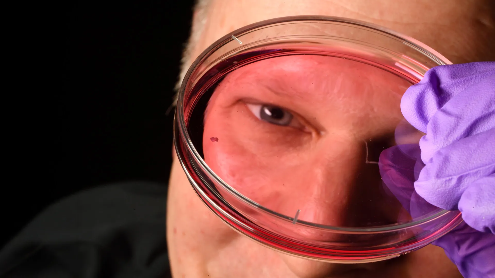
- Robert J. Johnston Jr., associate professor of biology at Johns Hopkins, led the study published in the Proceedings of the National Academy of Sciences, using retinal organoids grown from fetal cells and observed over several months.
- The foveola—a tiny region responsible for sharp central vision—contains only red and green cones yet accounts for about half of all human visual perception, and is the first region to fail in macular degeneration.
- The research could eventually enable cell replacement therapies for macular degeneration and glaucoma, with co-author Katarzyna A. Hussey—now at CiRC Biosciences—working to refine organoids that produce transplantable, healthy photoreceptors.
- Mice and fish, the most common lab animals, don't develop the same foveolar cone arrangement as humans, which is why the process has remained poorly understood for decades despite being central to human vision.
Why it matters: Johnston's team is now refining retinal organoids to produce healthier photoreceptors for transplant, targeting macular degeneration—a disease with no current cure that attacks the very foveola region the study decoded. The two-step retinoic acid–to–thyroid hormone sequence gives researchers a specific molecular timeline to engineer in the lab.
Ask SkimNews