Texas A&M Study Triggers Limb Regeneration in Mice — SkimNews
Get the Health newsletter
Daily health & science — research, biotech, public health, the studies worth knowing. Free.
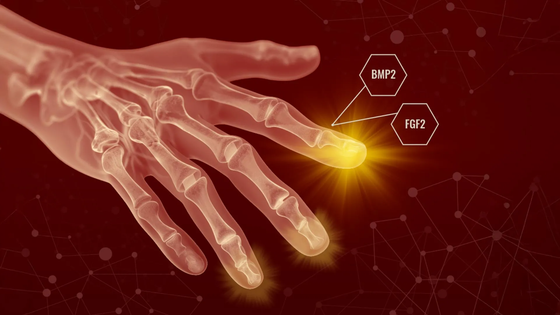
- Ken Muneoka and colleagues at Texas A&M's College of Veterinary Medicine and Biomedical Sciences published a study in Nature Communications describing a two-step treatment — FGF2 applied first, then BMP2 — that redirected healing from scar formation toward tissue regrowth after amputation in animal studies.
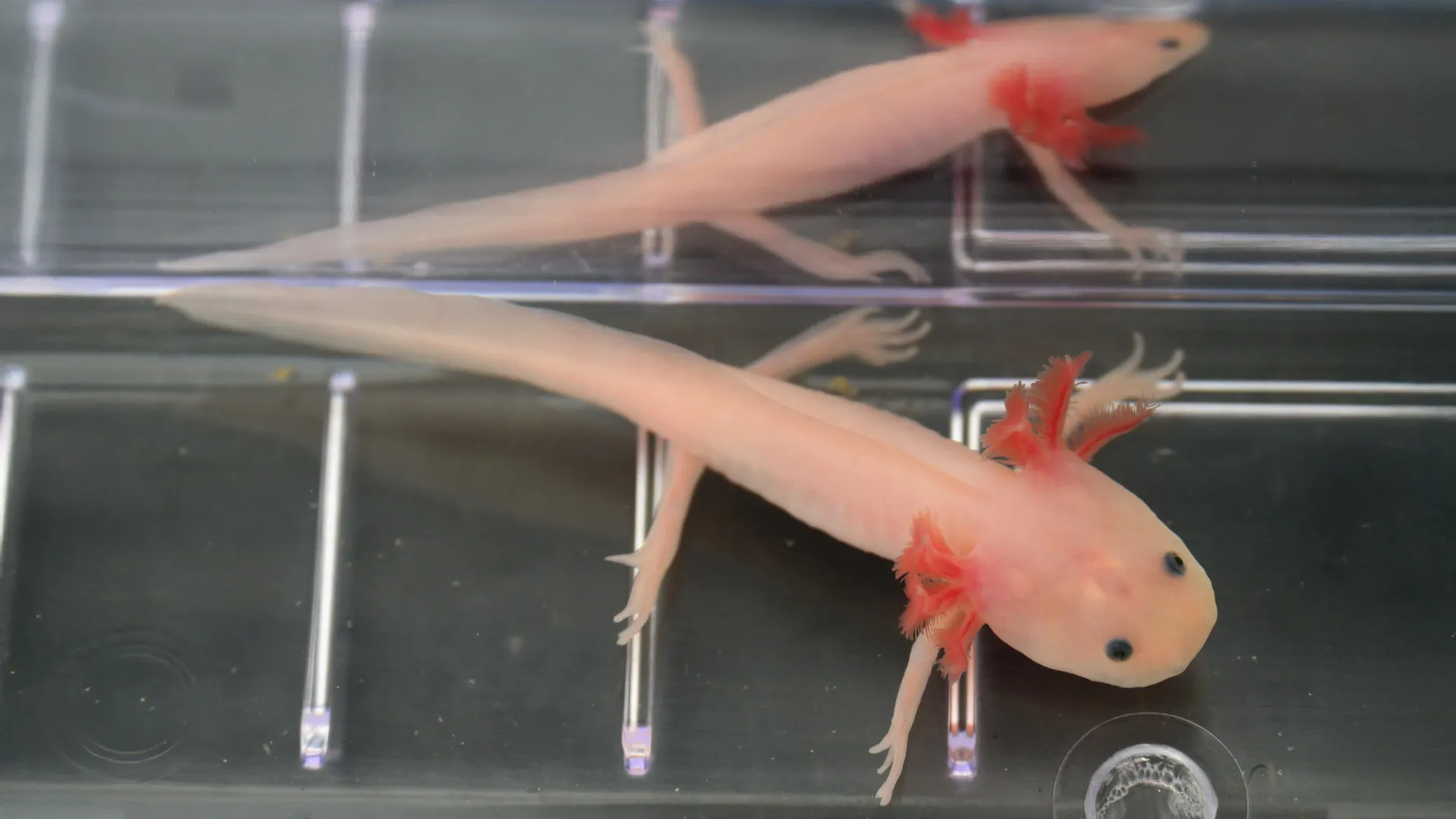
- The researchers regenerated bone, joints, ligaments, and tendons without adding external stem cells, instead reprogramming fibroblasts already present at the injury site into a blastema-like structure typically seen in salamanders.
- The regenerated tissues were not perfect replicas of the originals, but the major structures were present and arranged in patterns resembling natural anatomy, according to Muneoka.
- The study demonstrated positional re-specification — cells normally destined to form one tissue type were instructed to rebuild a different structure after injury, a process central to development.
- BMP2 already has FDA approval for certain medical applications, and FGF2 is being evaluated in multiple clinical trials, which the researchers say could shorten the path to human testing.
- Co-author Larry Suva said the findings overturn a long-standing assumption: "The cells that we thought to be unprogrammable, in fact are. The capacity is not absent — it's just obscured."
Why it matters: Regenerative medicine has long assumed mammals lack the cellular machinery to rebuild complex structures, making external stem cell delivery the dominant strategy. The Texas A&M team's finding that resident fibroblasts can be redirected — combined with BMP2's existing FDA approval — could compress the timeline from lab to clinic, offering amputees and trauma patients a path to regrown bone, tendon, and ligament tissue rather than scar.
Ask SkimNews