MIT Laser Pencil Beam Images Brain Barrier 25x Faster — SkimNews
Get the Health newsletter
Daily health & science — research, biotech, public health, the studies worth knowing. Free.
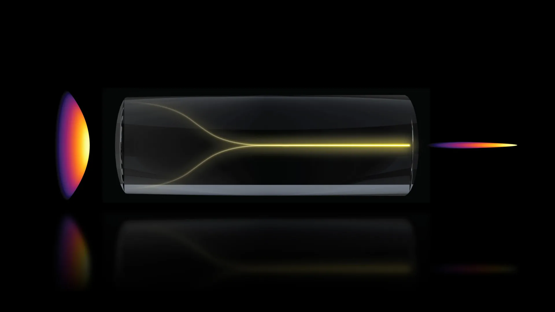
- MIT researchers found that increasing laser power inside a multimode optical fiber past the damage threshold made the light self-organize into a narrow, stable 'pencil beam'—contradicting the long-standing belief that high power inevitably produces chaotic, scattered light
- Sixian You, assistant professor in MIT's Department of Electrical Engineering and Computer Science, identified two required conditions: the laser must enter the fiber at a perfectly aligned zero-degree angle, and power must be high enough for the light to interact directly with the glass
- The team used the pencil beam to image the human blood-brain barrier in 3D roughly 25 times faster than the current gold-standard approach, while preserving comparable image quality and avoiding the resolution-versus-depth-of-focus tradeoff that limits conventional beams
- The technique can track how individual cells absorb drugs in real time without fluorescent labels—a first, according to co-author Roger Kamm—making it useful for screening Alzheimer's or ALS drug candidates on human-based tissue models where animal tests often fail to predict human outcomes
- The pencil beam lacks the blurred 'sidelobe' halos that degrade most comparable beams, keeping images clean and tightly focused across a large depth range
- The findings appear in Nature Methods, with first author Honghao Cao and collaborators from MIT, Harvard, and Beth Israel Deaconess Medical Center; the team plans to extend the method to imaging neurons and push toward practical deployment
- Funding came from MIT startup funds, the National Science Foundation, the Silicon Valley Community Foundation, the Diacomp Foundation, the Harvard Digestive Disease Core, a MathWorks Fellowship, and the Claude E. Shannon Award
Why it matters: Drug developers targeting the brain lose years when animal models fail to predict human blood-brain barrier permeability; a label-free imaging method that resolves cellular drug uptake in real time on human tissue could let pharmaceutical researchers screen candidates faster and catch failures earlier. The 25× speed gain, combined with no need for fluorescent tags, removes two of the biggest bottlenecks in live-tissue brain imaging.
Ask SkimNews