Laser Microscope Reveals Hidden Protein Machinery — SkimNews
Get the Health newsletter
Daily health & science — research, biotech, public health, the studies worth knowing. Free.
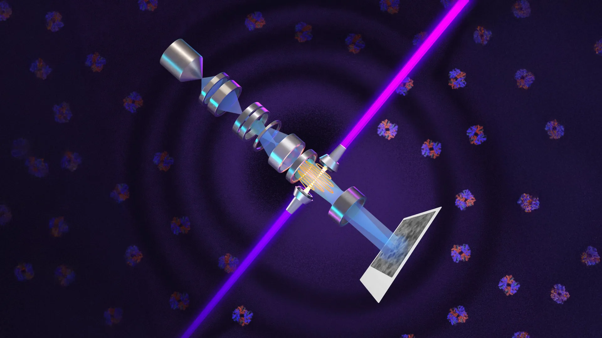
- University of California - Berkeley researchers created a laser phase plate that enhances electron microscopy by increasing image contrast through phase manipulation of the electron beam, allowing clearer visualization of tiny cellular structures.
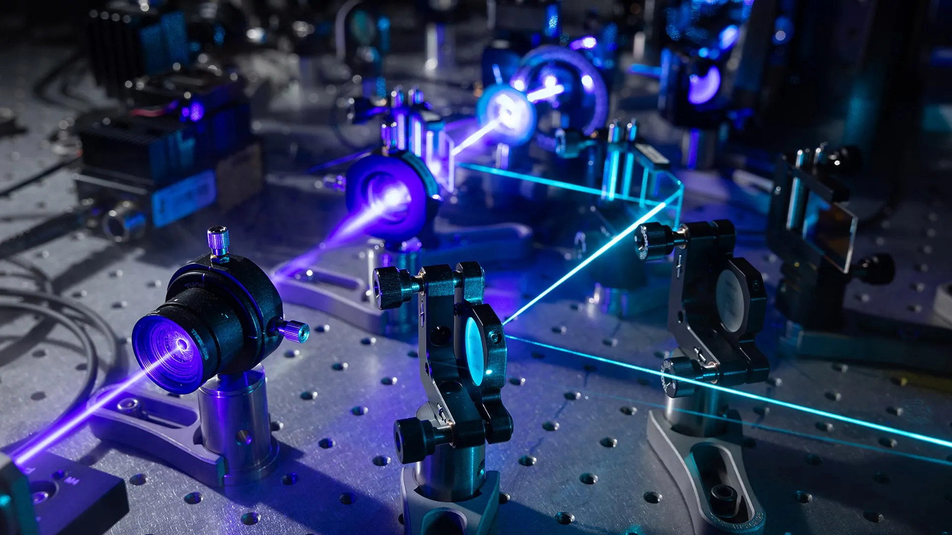
- Holger Müller led the development of a new microscope called Theia, which combines advanced electron optics with a high-intensity laser to achieve what he describes as potentially the world’s best resolution for biological imaging.
- Bridget Carragher is directing construction of a two-laser microscope at Biohub, building on Müller’s theoretical work, with the dual-laser setup designed to reduce optical distortions and prevent component damage.
- Theia uses the world’s most intense focused continuous-wave laser—75 kilowatts concentrated into a few microns—to shift the phase of electrons by 90 degrees, converting subtle cellular differences into visible contrast.
- Cryo-ET could gain the most from this technology, as it currently struggles with signal-to-noise limitations when imaging small proteins in crowded cellular environments, making molecular interactions hard to resolve.
- Thermo Fisher Scientific is collaborating with both the Berkeley and Biohub teams to integrate the laser phase plate technology into next-generation cryo-EM instruments, signaling industry recognition of its potential.
Why it matters: Scientists studying human proteins will gain access to clearer, more detailed 3D views of molecular machinery in their natural cellular context, overcoming a major signal-to-noise barrier that has limited cryo-EM and cryo-ET for years. This leap in imaging could accelerate drug discovery and structural biology research by making previously invisible targets visible.
Ask SkimNews