UC Berkeley Unveils Laser Microscope for Cellular Imaging — SkimNews
Get the Health newsletter
Daily health & science — research, biotech, public health, the studies worth knowing. Free.
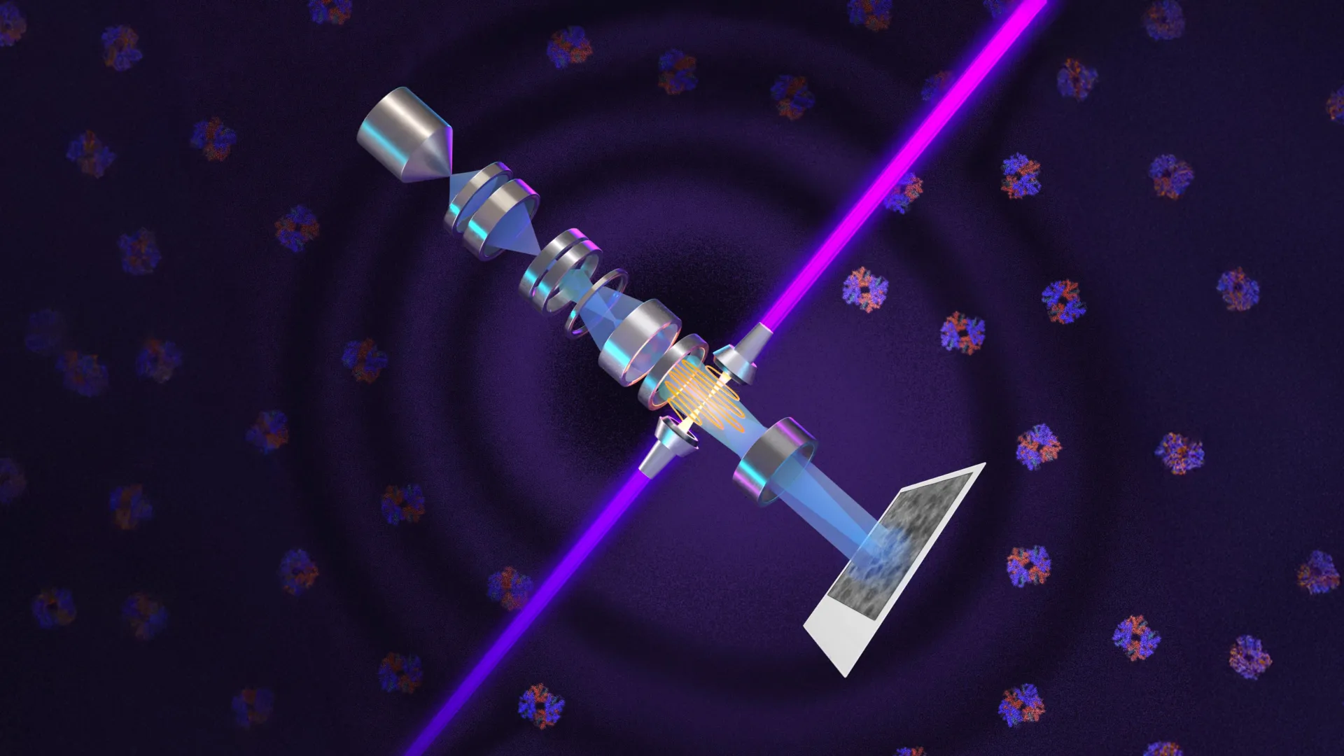
- University of California - Berkeley researchers created a laser phase plate that improves electron microscopy by increasing image contrast through phase manipulation of the electron beam, allowing clearer views of tiny cellular structures.
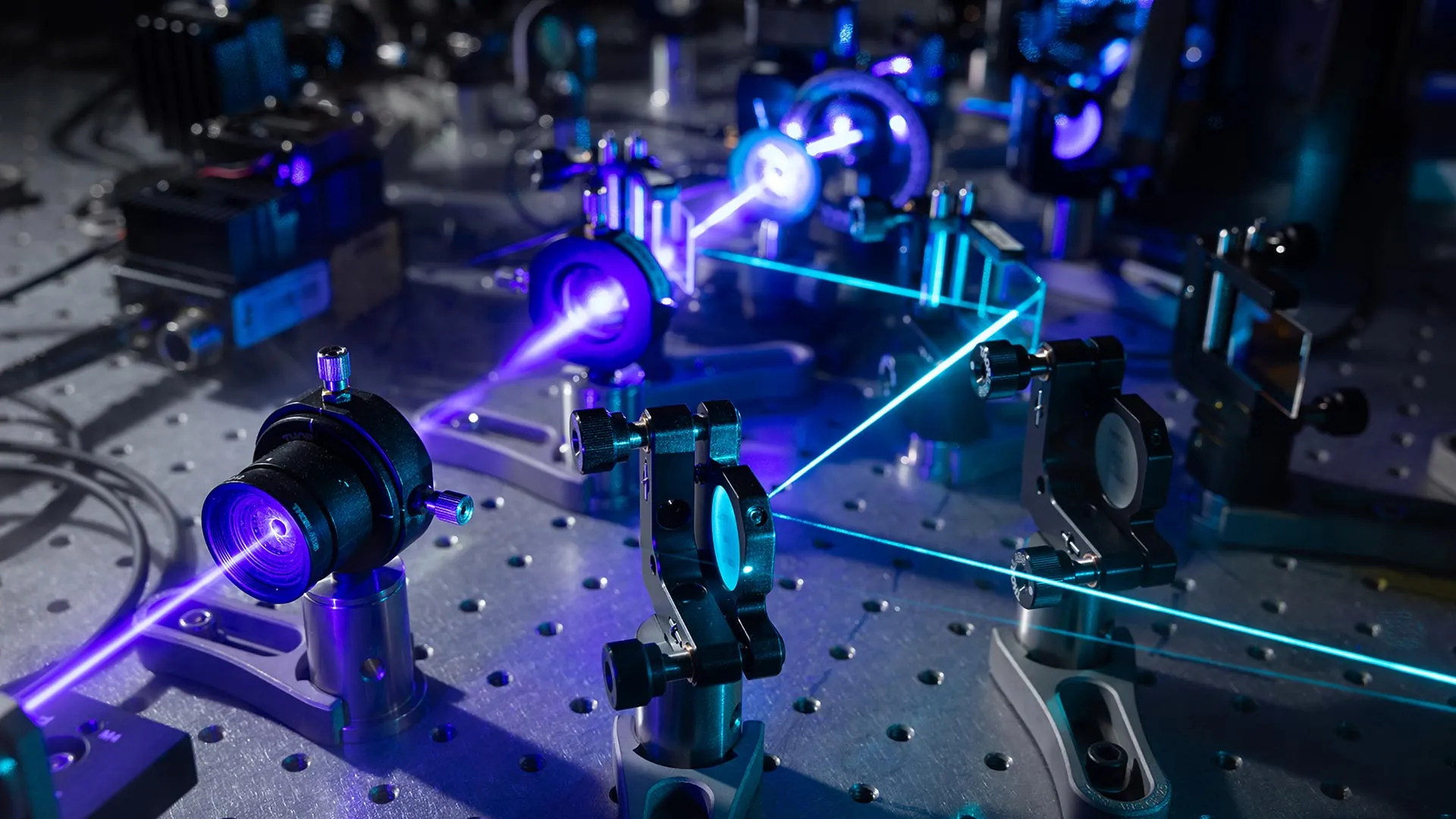
- Holger Müller led the development of a new microscope called Theia, which combines advanced electron optics with a high-intensity laser to achieve what he describes as potentially the world’s best resolution for biological imaging.
- Bridget Carragher is directing construction of a two-laser microscope at Biohub, based on Müller’s theoretical work, aiming to reduce optical distortions while maintaining high contrast for cryo-ET imaging.
- Theia uses the world’s most intense focused continuous-wave laser—75 kilowatts concentrated into a few microns—to shift the phase of electrons by 90 degrees, dramatically improving signal-to-noise ratio in cryo-EM.
- Cryo-ET could gain the most from this technology, as it currently struggles to image small proteins in crowded cellular environments; the laser phase plate promises to enhance contrast in 3D reconstructions of molecules in their natural context.
- Thermo Fisher Scientific is collaborating with both the Berkeley and Biohub teams, indicating industry engagement in advancing next-generation cryo-EM instrumentation.
Why it matters: Scientists studying molecular mechanisms of disease could now visualize proteins as small as one-third the size detectable by current cryo-EM, accelerating structural biology and drug discovery. The leap in imaging clarity reduces reliance on inference, making direct observation of cellular machinery feasible for the first time at this scale.
Ask SkimNews