USC: Midlife TV viewing linked to smaller brain regions — SkimNews

Get the Health newsletter
Daily health & science — research, biotech, public health, the studies worth knowing. Free.
- USC researchers published in Alzheimer's & Dementia a study of roughly 1,700 adults (average age 53) enrolled in the ARIC study between 1987 and 1989 who received brain MRI scans more than 20 years later.
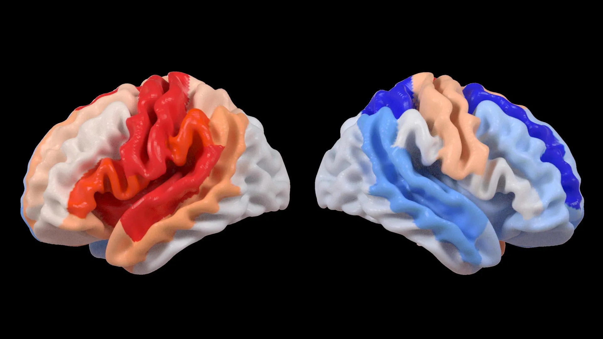
- Frequent midlife TV viewers showed smaller volumes in brain regions tied to memory, smaller frontal and occipital lobes, and greater white-matter hyperintensity volumes compared with people who reported watching "never/seldom".
- The associations held after researchers accounted for physical activity, diabetes, BMI, smoking, and alcohol use, according to the study.
- Workday sitting showed the opposite pattern: participants who spent long hours seated in mentally engaging jobs had larger frontal and occipital lobes and lower white-matter hyperintensity volumes, the study found.
- Brain changes were concentrated in men, with most TV- and sitting-linked structural differences appearing when MRI data was analyzed by sex.
- Lead authors David Raichlen and Natan Feter said the results suggest future guidance should focus on what people do while seated, not just total sitting time.
- Limitations noted: TV habits were self-reported, and no baseline MRI was taken at study entry, so researchers could not directly track structural change over time.
Why it matters: The finding flips the standard "sit less" prescription: in this cohort, mentally demanding seated work was tied to healthier brain structure than passive TV viewing, so any future public-health messaging has to distinguish between types of sedentary time. The data come from a 20-year longitudinal sample of ~1,700 adults, and the sex-specific effect (men drove most of the signal) means blanket dementia-prevention advice is unlikely to fit everyone equally.
Ask SkimNews