Max Planck Institute maps cancer sugar signatures — SkimNews
Get the Health newsletter
Daily health & science — research, biotech, public health, the studies worth knowing. Free.
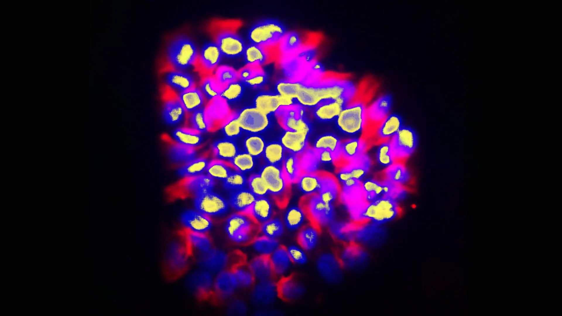
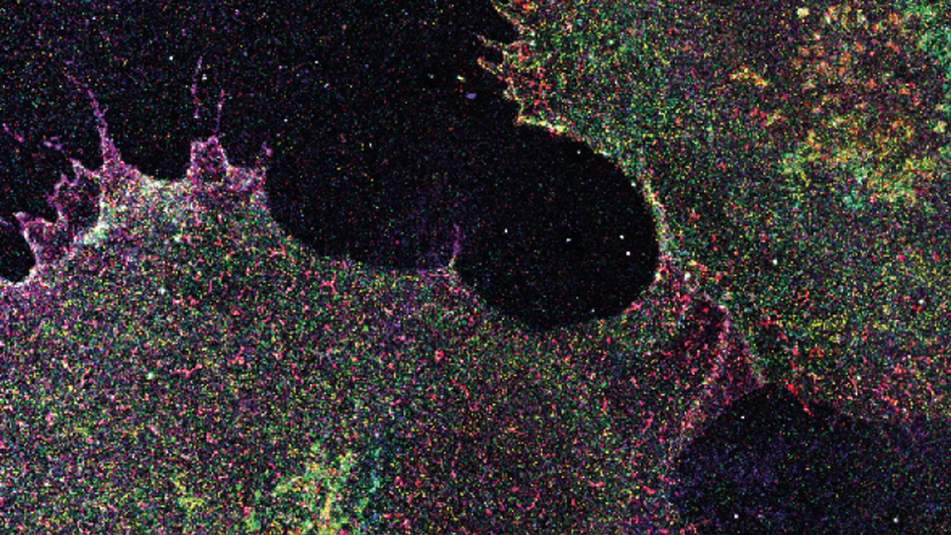
- Max Planck Institute for the Science of Light created a super‑resolution microscopy workflow named Glycan Atlasing to map individual sugar molecules on the glycocalyx of diverse human cells.
- Prof. Leonhard Möckl led the Physical Glycosciences group that applied Glycan Atlasing to cell lines, primary blood cells, and breast tissue samples.
- Immune cells displayed distinct glycocalyx sugar patterns after activation, showing that the surface sugar coating reflects functional state.
- Cancerous breast tissue showed a unique nanoscale sugar signature that could be distinguished from healthy tissue using the mapped glycocalyx patterns.
- Nature Nanotechnology published the study, underscoring the diagnostic potential of glycocalyx mapping for early disease detection.
Why it matters: Clinicians could gain a diagnostic tool that flags cancer earlier, potentially lowering treatment costs for hospitals, reducing the need for aggressive therapies, and improving patient survival rates.
Ask SkimNews