Cortisol Scrambles Brain's Internal GPS, MRI Study Finds — SkimNews
Get the Health newsletter
Daily health & science — research, biotech, public health, the studies worth knowing. Free.
- Ruhr-University Bochum researchers gave 40 healthy men 20mg of cortisol versus a placebo on separate days, then tested their navigation through a virtual meadow inside an MRI scanner; cortisol significantly increased orientation errors regardless of landmark availability or route complexity.
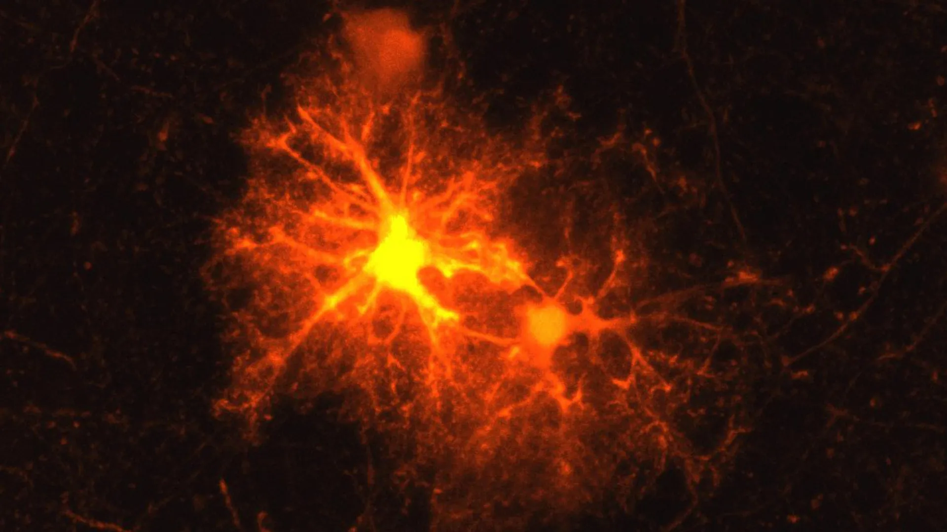
- Grid cells in the entorhinal cortex, which normally fire in precise repeating spatial patterns during navigation, showed much less distinct activity after cortisol — nearly vanishing when participants navigated without permanent landmarks.
- The caudate nucleus showed increased activity after cortisol dosing, indicating the brain attempted a compensatory navigation strategy when grid-cell mapping in the entorhinal cortex faltered.
- Dr. Osman Akan led the study with colleagues from Ruhr-University Bochum's Department of Cognitive Psychology and Neuropsychology plus University Hospital Hamburg-Eppendorf, and published the findings in PLOS Biology.
- The findings carry implications for Alzheimer's research, since the entorhinal cortex is among the first brain regions damaged by the disease and chronic stress is a known dementia risk factor.
Why it matters: This identifies a concrete mechanism linking cortisol to dysfunction in one of the earliest brain regions affected by Alzheimer's disease, giving dementia researchers a targetable pathway and giving chronic-stress sufferers a neurological explanation for why their sense of direction deteriorates.
Ask SkimNews