UCSF Team Finds Gut-Brain Pathway Behind Appetite Loss — SkimNews

Get the Health newsletter
Daily health & science — research, biotech, public health, the studies worth knowing. Free.
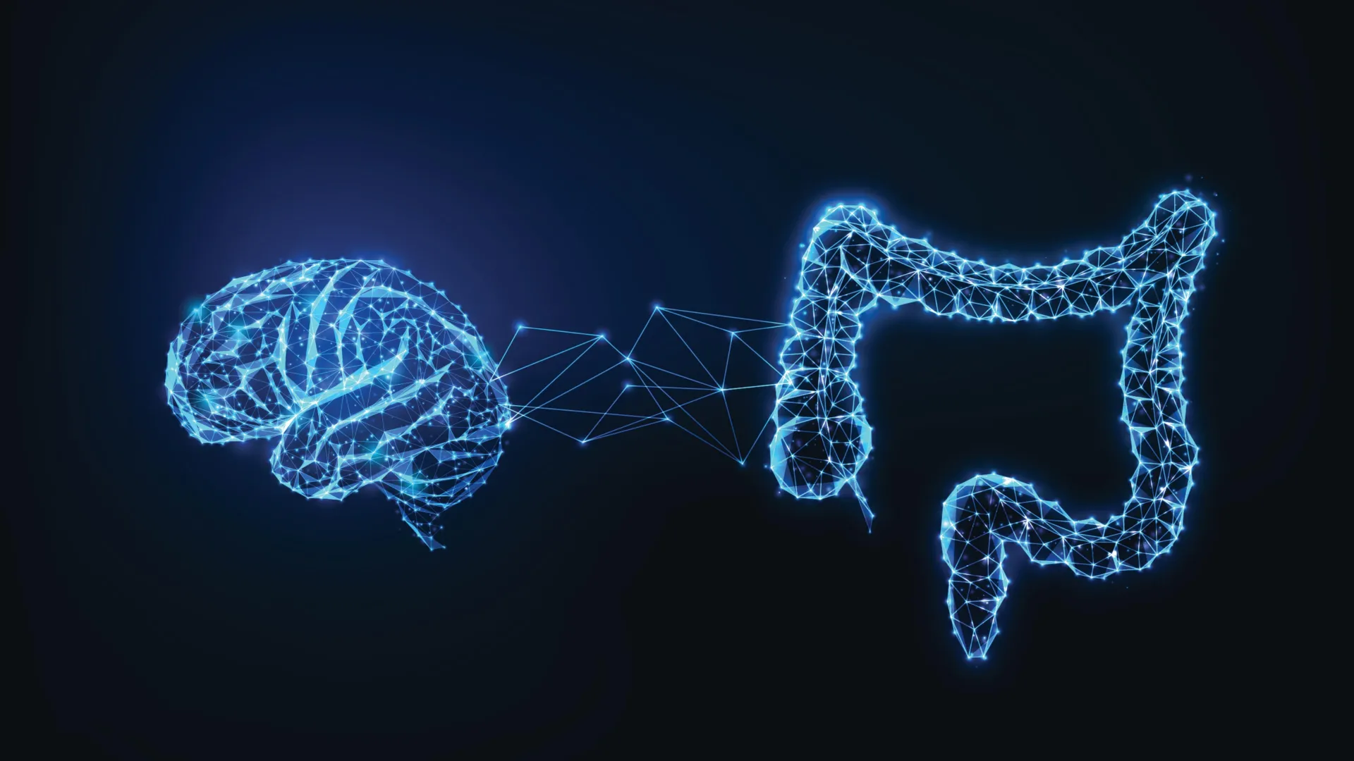
- UCSF researchers published a study in Nature on March 25, 2026, identifying how specialized gut cells called tuft cells detect parasites and ultimately suppress appetite in the brain.
- Tuft cells release acetylcholine when exposed to succinate from parasites, which triggers nearby enterochromaffin (EC) cells to release serotonin, sending signals to the brain via vagal nerve fibers.
- Tuft cells release acetylcholine in two phases — an initial short burst and a sustained release as the immune response builds — explaining why appetite loss is delayed rather than immediate after infection.
- David Julius, 2021 Nobel Prize laureate and co-senior author, said the gut "is essentially waiting to confirm that the threat is real and persistent" before telling the brain to change behavior.
- Mice with normal tuft cell function ate less as parasitic infection progressed, while mice unable to produce acetylcholine in their tuft cells continued eating normally — confirming the pathway drives the behavior.
- Researchers said the pathway's implications may extend beyond parasites to IBS, food intolerances, and chronic visceral pain, since tuft cells also exist in the airways, gallbladder, and reproductive system.
Why it matters: The study names a concrete molecular lever — acetylcholine release from tuft cells — that could eventually be modulated to treat appetite loss in the millions of people living with chronic parasitic infections, while reframing gut conditions like IBS and food intolerances as potential disorders of this same gut-brain signaling axis.
Ask SkimNews