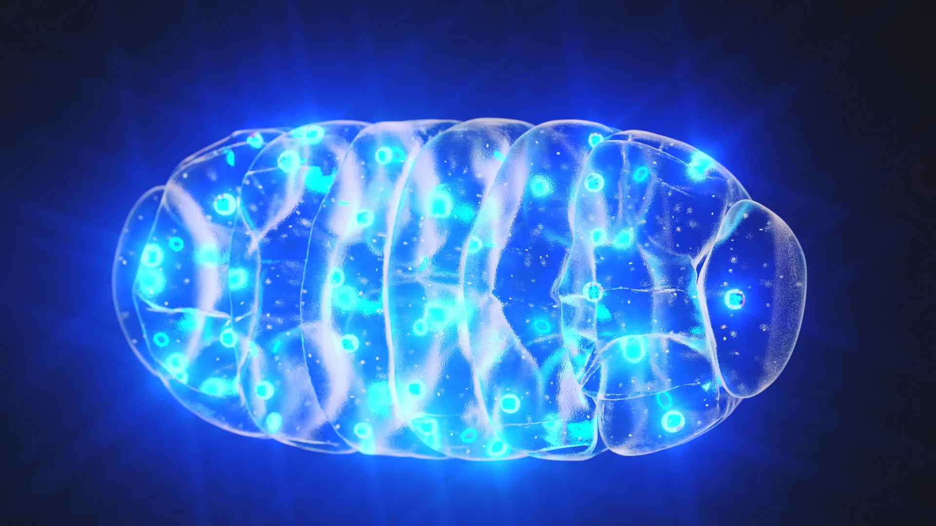
Göttingen Team Visualizes mHsp60 Stress Response — SkimNews

Get SkimNews daily
A curated daily digest across 7 categories: geopolitics, tech, finance, health, energy, sports, culture. Free.
- University Medical Center Göttingen researchers led by Prof. Dr. Rubén Fernández-Busnadiego used cryo-electron tomography to image flash-frozen human cells at approximately -200°C, preserving near-native cellular structures at near-atomic resolution.
- The team identified mHsp60 as a key folding helper that remodels its 3D structure under stress, forming barrel-like complexes that encapsulate and refold damaged mitochondrial proteins at increased rates.
- The study, published in Science Advances (DOI: 10.1126/sciadv.aed3579), was conducted with collaborators at the Biofisika Institute in Spain, Tel Aviv University in Israel, and the University of Dundee in the UK.
- Dopaminergic neurons in the midbrain, which demand high energy to produce dopamine and selectively degenerate in Parkinson's disease, are particularly vulnerable to mitochondrial dysfunction that the mHsp60 pathway counteracts.
- Parkinson's disease affects more than 10 million patients worldwide and has no cure, according to the umbrella organization Parkinson's Europe cited in the study.
- First author Kenneth Ehses, a postdoctoral researcher at UMG's Department of Neuropathology, said cryo-electron tomography enables studying protein complexes "directly within their native cellular environment" to investigate disease mechanisms.
- The team will next investigate how the mHsp60 folding cycle is altered under pathological conditions, including Parkinson's disease.
Why it matters: The technique lets researchers watch mitochondrial protein machinery operate inside actual cells, not test tubes, tying mHsp60's structural stress response directly to the dopaminergic neuron loss behind Parkinson's disease — a condition affecting more than 10 million patients worldwide with no existing cure, and one where drug developers have lacked a concrete molecular target until now.
Ask SkimNews