Axons form pearl-like chains, not smooth tubes — SkimNews
Get the Health newsletter
Daily health & science — research, biotech, public health, the studies worth knowing. Free.
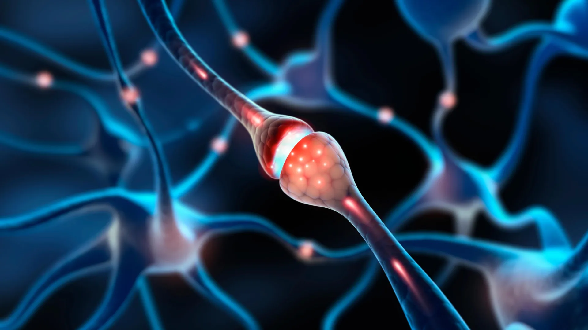
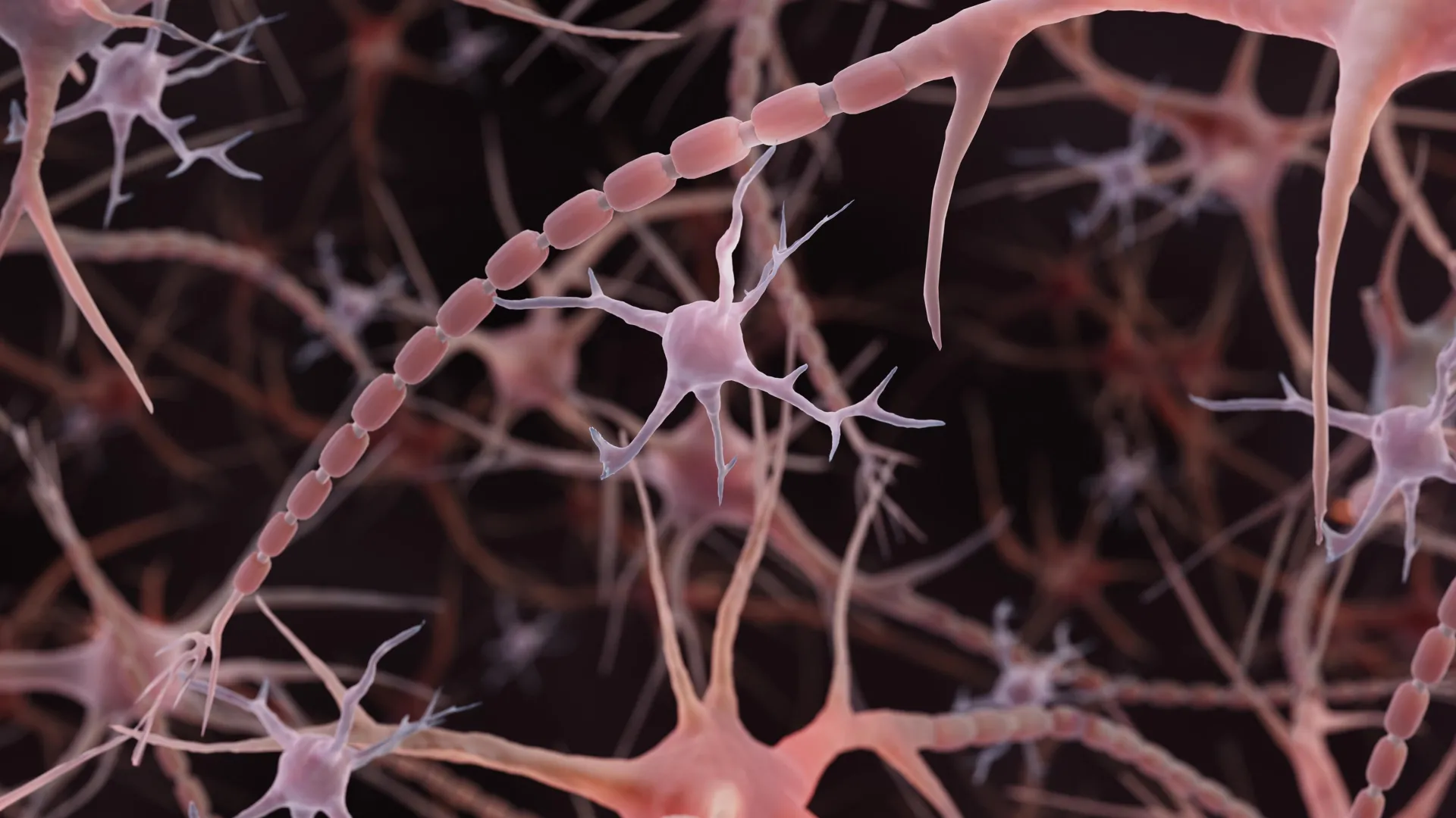
- Shigeki Watanabe and colleagues at Johns Hopkins found that mouse axons resemble strings of tiny pearls rather than the smooth tubes depicted in textbooks for more than a century, publishing in Nature Neuroscience on December 2, 2024.
- The team used high-pressure freezing electron microscopy — Watanabe compared it to freezing a grape instead of dehydrating it into a raisin — to preserve structures roughly 100 times narrower than a human hair.
- When disrupting the axon's internal protein skeleton failed to eliminate the pearls, the team partnered with UC San Diego biophysicist Padmini Rangamani, whose membrane-tension models reproduced the pearled pattern.
- High-frequency electrical stimulation made the pearl regions an average of 8% longer and 17% wider, and slowed electrical signals for at least an hour, the study reported.
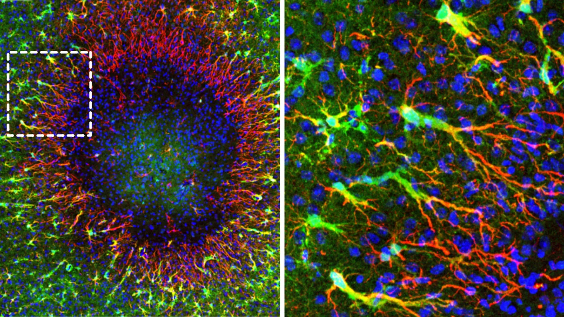
- A follow-up study in Neuron, led by Chelsy R. Eddings with Watanabe, documented pearled axons in human cortical tissue obtained during epilepsy surgery, extending the mouse observations.
- Removing cholesterol from the axon membrane made it more fluid, altered the pearled structure in both models and mouse neurons, and reduced electrical signaling speed.
Why it matters: With pearled axons now confirmed in human tissue and shown to respond to electrical activity, the finding gives neuroscience labs a new structural variable — membrane tension and composition — to investigate. The Johns Hopkins team explicitly flags an open question: distinguishing these normal activity-driven pearls from the damaging beading seen in Parkinson's and dying neurons, which could reshape how neurodegeneration is identified in tissue samples.
Ask SkimNews